Home
/ Cells Under Microscope - Onion Cells Under A Microscope 400x 1000x Youtube - Some have been increased up to 5,000 times!
Cells Under Microscope - Onion Cells Under A Microscope 400x 1000x Youtube - Some have been increased up to 5,000 times!
Cells Under Microscope - Onion Cells Under A Microscope 400x 1000x Youtube - Some have been increased up to 5,000 times!. What is the length of an onion cell indicated by the pointer in micrometers? After the mrna vaccine, the images of the red blood cells have an abnormal membrane and start to clump together. A cuboidal epithelial cell looks close to a square. The revelation was pretty shocking. The cell membrane acts like a balloon and holds all the parts of a cell inside, such as a nucleus, cytosol, and organelles.
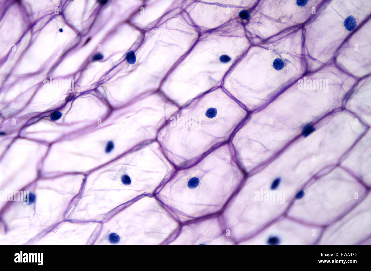
We measured the cell size by using a microscopic meter slide. After you get the mrna vaccine, the membrane of red blood cells becomes abnormal and they clump together. A columnar epithelial cell looks like a column or a tall rectangle. Below we have put together a list of images of different parts of the human body under the microscope. While photosynthesis takes place in the leaves of an onion containing chloroplast, the little glucose that is produced from this process is converted in to starch (starch granules) and stored in the bulb.
Human Blood Cells Under Microscope View Stock Photo Picture And Royalty Free Image Image 81293536 from previews.123rf.com The red stuff we see is lipids stored within the membrane of each individual cell, ready for use when needed. Now we know what happens when jabs enter your bodies. Observing cancer cells under the microscope one of the more useful and essential uses of microscopy is in identifying, analyzing, and treating certain diseases, ranging anywhere from bacterial and viral infections, to something a lot more serious and fatal, such as cancer. See cell under microscope stock video clips. A squamous epithelial cell looks flat under a microscope. Peel a translucent piece of tissue from the onion. A cuboidal epithelial cell looks close to a square. Students will observe cheek cells under a microscope.
Use them in commercial designs under lifetime, perpetual & worldwide rights.
Recently, images of red blood cells under the microscope have appeared, showing some shocking revelations of what the vaccine does to the body. While photosynthesis takes place in the leaves of an onion containing chloroplast, the little glucose that is produced from this process is converted in to starch (starch granules) and stored in the bulb. University of california, san francisco We are avid microscope enthusiasts and general. Now we know what happens when jabs enter your bodies. Create custom image collections with your shutterstock account. What is the length of an onion cell indicated by the pointer in micrometers? Observing cancer cells under the microscope one of the more useful and essential uses of microscopy is in identifying, analyzing, and treating certain diseases, ranging anywhere from bacterial and viral infections, to something a lot more serious and fatal, such as cancer. Types of cells that can be viewed under a basic compound microscope include cork cells, plant cells and even human cells scraped from the inside of the cheek. Dreamstime is the world`s largest stock photography community. Many items at sale prices. After you get the mrna vaccine, the membrane of red blood cells becomes abnormal and they clump together. They are among the largest cells in our bodies (as many of us are painfully aware).
Red blood cells are responsible for transporting oxygen throughout the body. Onion cells under a microscope requirements, preparation and observation the bulb of an onion is formed from modified leaves. Below we have put together a list of images of different parts of the human body under the microscope. How many cells should be visible in low power (10x) field of view? How many cells are visible in this high power (40 x) field of view?
Blue Organic Cells Under Microscope Stock Photo Alamy from c8.alamy.com Below is a size and length scale in biology, including eggs, cells, organelles, bacteria, viruses, protein complexes, and atoms. Everything can look strange under a microscope, even the human body. And that's only the beginning. Red blood cells (rbcs) as seen under the microscope in isotonic, hypotonic and hypertonic solutions. How many cells should be visible in low power (10x) field of view? Microglial cells recognize areas of damage and inflammation and swallow cellular debris. Find professional human cells under microscope videos and stock footage available for license in film, television, advertising and corporate uses. I did this by lowering the condenser lens and closing the iris diaphragm to match the 0.65 numerical aperture of the 40x objective i was using.
Peel a translucent piece of tissue from the onion.
We measured the cell size by using a microscopic meter slide. This check cell is about 80 micrometers in diameter. Many items at sale prices. Find out how to observe cells under a microscope click to. When we look at cells under the microscope, our usual measurements fail to work. What is the procedure of viewing cells? Use them in commercial designs under lifetime, perpetual & worldwide rights. Best way to observe blood under the microscope to see the distinctive red blood cell disk shape, you need a little bit of contrast. After you get the mrna vaccine, the membrane of red blood cells becomes abnormal and they clump together. Monthly plans starting at $29. A squamous epithelial cell looks flat under a microscope. How many cells are visible in this high power (40 x) field of view? What is the length of an onion cell indicated by the pointer in micrometers?
Human cells under a microscope stem cells background human cells under microscope molecular cell. Assume that the onion cells photo was taken under the high power field of microscope from question 4. Find out how to observe cells under a microscope click to. After the mrna vaccine, the images of the red blood cells have an abnormal membrane and start to clump together. Monthly plans starting at $29.
Onion Epidermis With Large Cells Under Light Microscope Clear Epidermal Cells Of An Onion Allium Cepa In A Single Layer Stock Photo Alamy from c8.alamy.com After the mrna vaccine, the images of the red blood cells have an abnormal membrane and start to clump together. The green splash is a microglial cell, which responds to immune reactions in the central nervous system. (the smaller the piece the better.) translucent means that you can see light through the specimen, but. What is the length of an onion cell indicated by the pointer in micrometers? When we look at cells under the microscope, our usual measurements fail to work. The larger orange shape is an oligodendrocyte. While photosynthesis takes place in the leaves of an onion containing chloroplast, the little glucose that is produced from this process is converted in to starch (starch granules) and stored in the bulb. Below we have put together a list of images of different parts of the human body under the microscope.
They are among the largest cells in our bodies (as many of us are painfully aware).
Create custom image collections with your shutterstock account. We are avid microscope enthusiasts and general. After the mrna vaccine, the images of the red blood cells have an abnormal membrane and start to clump together. Cheek cells under a microscope requirements, preparation and staining cheek cells are eukaryotic cells (cells that contain a nucleus and other organelles within enclosed in a membrane) that are easily shed from the mouth lining. Under the microscope, animal cells appear different based on the type of the cell. A few epithelial layers are constructed from cells that are said to have a transitional shape. Find out how to observe cells under a microscope click to. Onion cells under a microscope requirements, preparation and observation the bulb of an onion is formed from modified leaves. Microglial cells recognize areas of damage and inflammation and swallow cellular debris. A few white blood cells can also be seen with the red bl. Monthly plans starting at $29. A single yeast cell is typically about 3 to 4 micrometers in diameter but there are some species that can grow to be much larger, around 40 micrometers. Your cells under microscope stock images are ready.